
Nucleic acids are the molecules of life — they store, transmit, and express genetic information. Without DNA and RNA, cells would lack instructions for protein synthesis, inheritance, and regulation of cellular processes. This unit explores the biosynthesis and breakdown of nucleotides, organization of the genome, replication, transcription, and translation, highlighting how these processes ensure life continues from one generation to the next.
Biosynthesis of Purine and Pyrimidine Nucleotides
Bioenergetics is the study of energy transfer and transformation in living organisms. It’s a fundamental field of biochemistry that focuses on how cells acquire, convert, and use energy to perform biological work. The core principles of bioenergetics are based on the laws of thermodynamics, which govern all energy transfer.
Key Principles
- First Law of Thermodynamics: Energy cannot be created or destroyed, only converted from one form to another. In living systems, this means the chemical energy in food is converted into other forms of energy (e.g., kinetic energy for movement, thermal energy as heat).
- Second Law of Thermodynamics: In any energy conversion, some energy is lost as heat, leading to an increase in the entropy (disorder) of the universe. This is why metabolic processes are not 100% efficient.
Metabolic Processes
Bioenergetics is primarily concerned with the two major metabolic pathways that manage energy:
- Catabolism: The breakdown of complex molecules into simpler ones, releasing energy in the process. This is an exergonic reaction. For example, the catabolism of glucose through cellular respiration releases energy to be stored as ATP.
- Anabolism: The synthesis of complex molecules from simpler ones, which requires an input of energy. This is an endergonic reaction. For example, the synthesis of proteins from amino acids or the storage of glucose as glycogen.
Catabolism of Purine Nucleotides and Disorders
The catabolism of purine nucleotides is the metabolic pathway that breaks down purine bases (adenine and guanine) into a final product for excretion. Disorders in this pathway can lead to various medical conditions due to the accumulation or deficiency of its intermediates.
Catabolism of Purine Nucleotides
The breakdown of purine nucleotides occurs in a series of enzymatic steps.
- AMP Breakdown:
- Adenosine monophosphate (AMP) is dephosphorylated to form adenosine.
- Adenosine is then deaminated by adenosine deaminase to form inosine.
- Inosine is broken down to hypoxanthine.
- Hypoxanthine is oxidized by the enzyme xanthine oxidase to form xanthine.
- Xanthine is then further oxidized by the same enzyme, xanthine oxidase, to form uric acid.
- GMP Breakdown:
- Guanosine monophosphate (GMP) is dephosphorylated to form guanosine.
- Guanosine is broken down to guanine.
- Guanine is deaminated to form xanthine.
- As with AMP catabolism, xanthine is oxidized by xanthine oxidase to form uric acid.
Uric acid is the final end product of purine catabolism in humans. It is transported to the kidneys and excreted in the urine.
Disorders of Purine Catabolism
Disorders of this pathway are often caused by genetic defects in the enzymes involved.
- Gout: This is the most common disorder related to purine catabolism. It is caused by the overproduction of uric acid or the under-excretion of it by the kidneys, leading to hyperuricemia (high uric acid levels in the blood). The excess uric acid crystallizes in the joints, causing painful inflammation and arthritis.
- Lesch-Nyhan Syndrome (LNS): A rare, severe, X-linked genetic disorder caused by a deficiency in the enzyme hypoxanthine-guanine phosphoribosyltransferase (HGPRT). This deficiency leads to a buildup of hypoxanthine and guanine, which are then shunted to the uric acid pathway, causing severe hyperuricemia. LNS is characterized by intellectual disability, spasticity, self-mutilating behavior, and gout-like symptoms.
- Severe Combined Immunodeficiency (SCID): One form of SCID is caused by a deficiency in the enzyme adenosine deaminase (ADA). The lack of this enzyme leads to the accumulation of dATP, which is toxic to developing lymphocytes (B and T cells). This toxicity severely impairs the immune system, making individuals highly susceptible to infections.
Organization of the Mammalian Genome
The mammalian genome is organized into a highly intricate and compact structure. This organization allows the vast amount of genetic information to be stored within the nucleus of a cell and be accessible for processes like gene expression and replication. The organization of the genome is hierarchical, moving from the basic DNA double helix to the condensed chromosome.
Levels of Organization
- DNA Double Helix: The fundamental level is the DNA molecule itself, a double helix composed of two strands held together by hydrogen bonds. It consists of a sequence of four nucleotides: adenine (A), thymine (T), guanine (G), and cytosine (C).
- Nucleosomes: The DNA double helix is wrapped around a core of eight proteins called histones. This DNA-histone complex is known as a nucleosome. This winding compacts the DNA by a factor of about 7.
- Chromatin Fiber: The nucleosomes are then coiled and stacked to form a thicker, more condensed structure called a 30 nm chromatin fiber. This fiber is further organized into loops and domains.
- Chromatid and Chromosomes: The chromatin fiber is folded and compacted into a larger structure called a chromatid. During cell division (mitosis or meiosis), two identical chromatids are joined at a centromere to form a chromosome, the most condensed form of the genome.
Structure of DNA and RNA
DNA and RNA are nucleic acids, which are complex organic molecules essential for life. Both are polymers made of repeating units called nucleotides, but they have key differences in their structure and function.
The Basic Unit: Nucleotides
A nucleotide consists of three main components:
- A Pentose Sugar: A five-carbon sugar.
- A Phosphate Group: A negatively charged group.
- A Nitrogenous Base: A ring-structured molecule containing nitrogen.
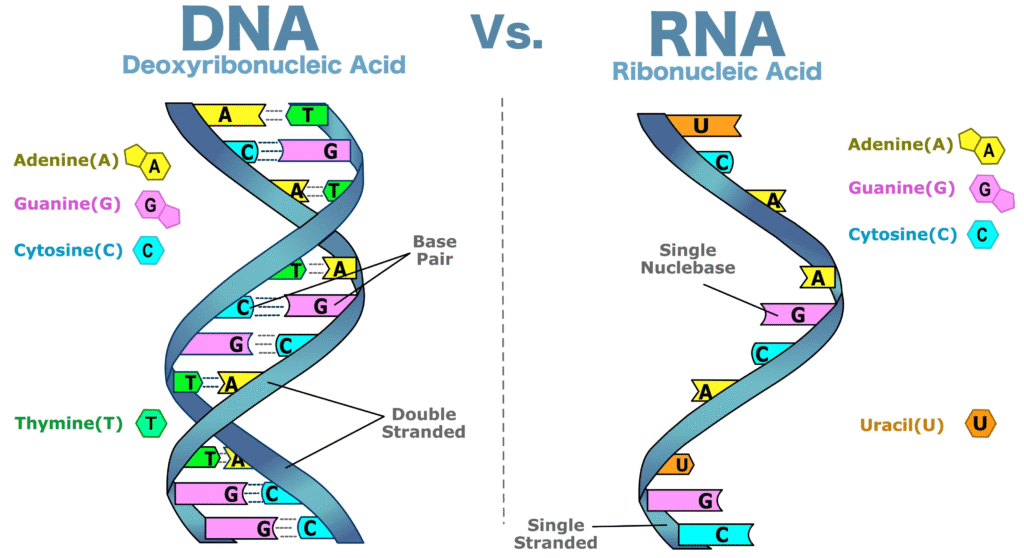
DNA (Deoxyribonucleic Acid)
DNA is the genetic blueprint of an organism. Its structure is a double helix, resembling a twisted ladder.
- Sugar: The sugar in DNA is deoxyribose, which has one less oxygen atom than ribose.
- Bases: DNA contains four bases: adenine (A), guanine (G), cytosine (C), and thymine (T).
- Strands: DNA is made of two long polynucleotide strands that run in opposite directions (antiparallel). The sugar-phosphate backbone forms the sides of the ladder, and the nitrogenous bases form the rungs.
- Base Pairing: The two strands are held together by hydrogen bonds between the bases. This pairing is highly specific: adenine always pairs with thymine (A-T), and guanine always pairs with cytosine (G-C).
RNA (Ribonucleic Acid)
RNA is primarily involved in the synthesis of proteins. Its structure is typically a single strand, though it can fold into complex 3D shapes.
- Sugar: The sugar in RNA is ribose.
- Bases: RNA contains four bases: adenine (A), guanine (G), cytosine (C), and uracil (U). Uracil replaces thymine found in DNA.
- Strand: RNA is usually a single polynucleotide strand.
- Base Pairing: Although single-stranded, RNA can form temporary, localized double-stranded regions through complementary base pairing (A-U and G-C), allowing it to fold into functional structures.