
The nervous system is one of the most complex and vital systems of the human body. It acts like a command and communication network, controlling our thoughts, emotions, movements, and responses to the environment. By coordinating information between the body and brain, it maintains homeostasis and ensures survival.
This unit introduces the organization of the nervous system, the structure and function of neurons and supporting cells, the physiology of nerve conduction, as well as the detailed anatomy of the central nervous system (CNS).
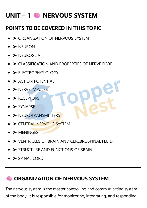
Organization of the Nervous System
The nervous system is organized into two main parts: the central nervous system (CNS) and the peripheral nervous system (PNS). This division helps to understand how information is processed and communicated throughout the body.
Central Nervous System (CNS)
The CNS is the main control center of the body. It consists of the brain and the spinal cord. Its primary function is to process, interpret, and store incoming sensory information and to send out instructions to the rest of the body.
- Brain: The brain is the seat of consciousness, thought, memory, and emotion. It integrates sensory information and coordinates all bodily activities.
- Spinal Cord: The spinal cord is a long column of nervous tissue that extends from the brain. It serves as a communication highway between the brain and the body, and it also controls simple, involuntary reflexes without needing input from the brain.
Peripheral Nervous System (PNS)
The PNS consists of all the nerves that lie outside the CNS. Its main role is to connect the CNS to the limbs and organs, acting as a communication network. It is further divided into two main branches:
- Somatic Nervous System: This system controls voluntary movements and transmits sensory information from the skin and muscles to the CNS. It is responsible for actions you consciously control, like walking or writing.
- Autonomic Nervous System (ANS): The ANS controls involuntary functions like heart rate, breathing, and digestion. It operates without conscious thought. The ANS is further divided into two opposing systems:
- Sympathetic Nervous System: This is the “fight-or-flight” response system. It prepares the body for action by increasing heart rate, dilating pupils, and slowing down digestion.
- Parasympathetic Nervous System: This is the “rest-and-digest” system. It calms the body down after a stressful event, lowering heart rate and stimulating digestion.
Neuron – The Structural and Functional Unit
A neuron is the basic structural and functional unit of the nervous system. These specialized cells are responsible for transmitting electrical and chemical signals, allowing for communication between different parts of the body. They are the core components of the brain, spinal cord, and peripheral nerves.
Structure of a Neuron
A typical neuron is composed of three main parts:
- Dendrites: These are short, branching extensions that receive incoming signals from other neurons. They act like antennae, gathering information and transmitting it toward the cell body. A single neuron can have many dendrites.
- Cell Body (Soma): This is the main part of the neuron, containing the nucleus and other essential organelles. It integrates the signals received from the dendrites and generates a new signal to be transmitted down the axon.
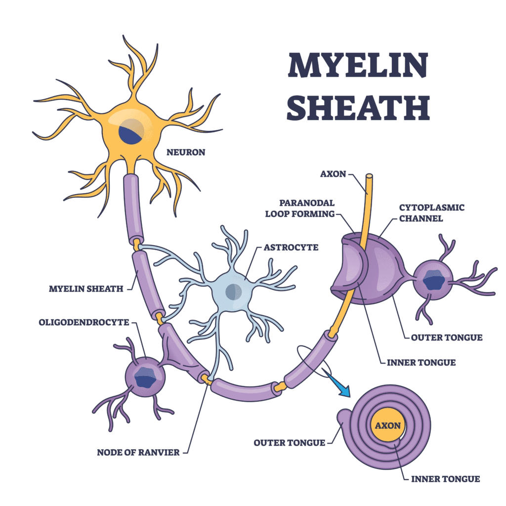
- Axon: A long, slender projection that extends from the cell body and transmits electrical impulses away from the cell. The axon is often covered in a myelin sheath, a fatty layer that insulates the axon and speeds up the transmission of the nerve impulse. The axon ends in a series of branching terminals that connect to other neurons or target cells.
Receptors
Receptors are protein molecules, typically located on the surface of cells or within the cytoplasm, that bind to specific signaling molecules (ligands). This binding event triggers a cascade of internal changes within the cell, leading to a biological response. In pharmacology, the ligands are often drugs, hormones, or neurotransmitters, and the response is the therapeutic effect or side effect of the drug.
Key Characteristics of Receptors
- Specificity: Receptors are highly specific. A particular receptor will only bind to a specific type of ligand or a class of chemically similar ligands. This is often compared to a “lock and key” mechanism. For example, a receptor for the hormone insulin will not bind to the hormone adrenaline.
- Affinity: This refers to the strength of the bond between a ligand and its receptor. A drug with a high affinity will bind tightly to its receptor, even at low concentrations.
- Saturability: There is a finite number of receptors on a cell. Once all the available receptors are bound to ligands, the system is saturated, and increasing the ligand concentration will not increase the response.
- Reversibility: The binding of a ligand to a receptor is generally reversible. The ligand can bind and unbind from the receptor, which allows the cellular response to be transient and regulated.
Types of Receptors
Receptors are classified based on their location and the type of signal they transmit.
- Ion Channel-Linked Receptors: These are transmembrane proteins that form an ion channel. When a ligand binds to the receptor, the channel opens, allowing ions to flow across the membrane and alter the cell’s electrical potential. These are involved in fast synaptic transmission.
- G-Protein Coupled Receptors (GPCRs): These are the largest family of cell-surface receptors. When a ligand binds, it activates an associated G-protein, which then triggers a cascade of intracellular signaling events. They are involved in a wide variety of physiological processes.
- Enzyme-Linked Receptors: These are transmembrane proteins with an intracellular enzymatic domain. Ligand binding activates the enzyme, which then catalyzes a reaction inside the cell.
- Intracellular Receptors: These receptors are located inside the cytoplasm or nucleus. The ligands for these receptors are typically small, lipid-soluble molecules (like steroid hormones) that can pass through the cell membrane. The ligand-receptor complex then moves to the nucleus to directly influence gene expression.
Introduction to Central Nervous System (CNS)
The Central Nervous System (CNS) is the main control center of the body. It consists of the brain and the spinal cord. Its primary function is to process, interpret, and store incoming sensory information and to send out instructions to the rest of the body.
Structure & Functions of the Brain
The brain is a complex organ that serves as the command center of the nervous system. It is responsible for processing, interpreting, and integrating information from the body and the external environment.
Major Structures of the Brain
The brain is divided into three main parts: the forebrain, the midbrain, and the hindbrain.
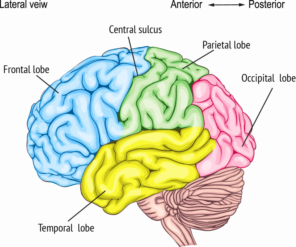
- Forebrain: The largest and most complex part of the brain. It contains the cerebrum, which is responsible for higher-level functions like thought, language, and voluntary actions. The cerebrum is divided into two hemispheres and four lobes:
- Frontal Lobe: Controls reasoning, planning, movement, and problem-solving.
- Parietal Lobe: Processes sensory information like touch, temperature, and pain.
- Occipital Lobe: Processes visual information.
- Temporal Lobe: Processes auditory information, memory, and speech.
- Midbrain: Located between the forebrain and the hindbrain, the midbrain is a small but vital part of the brain stem. It coordinates movement and is involved in processing auditory and visual information.
- Hindbrain: Located at the back of the head, the hindbrain consists of the cerebellum, the pons, and the medulla oblongata.
- Cerebellum: Coordinates voluntary movements, maintains balance, and regulates posture.
- Pons: Acts as a bridge, relaying signals between the cerebrum and the cerebellum. It is also involved in sleep and breathing.
- Medulla Oblongata: The lowest part of the brainstem, it controls involuntary functions vital for survival, such as heart rate, breathing, and blood pressure.
Spinal Cord
The spinal cord is a long, tubular bundle of nervous tissue that extends from the base of the brain down the center of the back. It serves as a vital communication highway between the brain and the rest of the body.
Structure of the Spinal Cord
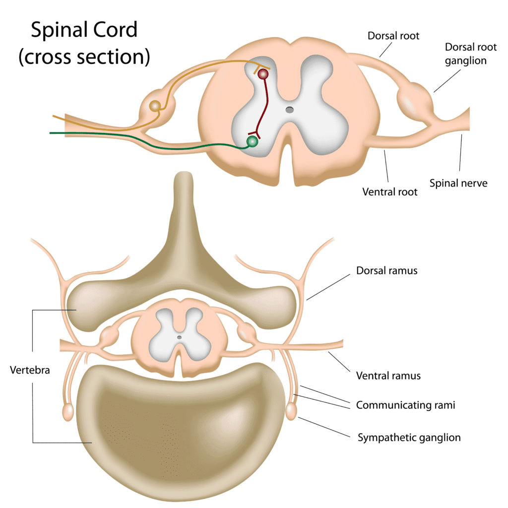
The spinal cord is housed within the spinal column, which provides bony protection. It is a cylinder of tissue with two main regions when viewed in a cross-section:
- Gray Matter: Located in the center, this region is shaped like a butterfly or an “H.” It contains the cell bodies of neurons, dendrites, and glial cells. The gray matter is where information is processed and where reflexes are initiated.
- White Matter: Surrounding the gray matter, this region is composed of myelinated axons. The white matter acts as the communication lines, with nerve fibers (tracts) that run up and down the spinal cord, carrying signals to and from the brain.